Airway hyperresponsiveness in severe asthma

Airway hyperresponsiveness results from an intricate interplay of airway inflammation, airway remodelling and structural changes.1–3

Airway hyperresponsiveness is a cardinal feature of asthma1

  • Airway hyperresponsiveness is the heightened bronchoconstrictive response to stimuli that would produce little or no effect in healthy individuals1,2 ​
  • Airway hyperresponsiveness is considered to be a key feature in the diagnosis, classification of severity and management of asthma, and is associated with various asthma symptoms including wheezing, chest tightness and bronchoconstriction1,3
  • Airway hyperresponsiveness results from an intricate interplay of airway inflammation, airway remodelling ​and structural changes;1,2,4 infiltration of airway smooth muscle by mast cells, and the resultant secretion of ​pro-inflammatory and profibrotic mediators, plays a major role in airway hyperresponsiveness5​
  • Airway hyperresponsiveness is dynamic; its magnitude and presence can vary over time with disease activity, specific exposures and with treatment, and the severity of airway hyperresponsiveness is also associated with increased asthma severity1,2,6

Airway hyperresponsiveness is suggested to be a ‘treatable trait’ that could be managed by precision medicine in patients with severe asthma7

1. Chapman DG, Irvin CG. Clin Exp Allergy 2015;45:706–719; 2. Comberiati P, et al. Immunol Allergy Clin North Am 2018;38:545–571; 3. Rayees S, Din I. Asthma: pathophysiology, ​herbal and modern therapeutic interventions. Cham, Switzerland: Springer International Publishing, 2021; 4. Berair R, et al. J Allergy (Cairo) 2013;2013:185971; 5. Bradding P. Eur Respir J 2007;29:827–830; ​6. in’t Veen JC, et al. Am J Respir Crit Care Med 1999;160:93–99; 7. Charriot J, et al. Eur Resp J 2023;61:2202389

What is airway hyperresponsiveness?

Airway hyperresponsiveness is a cardinal feature of asthma.1 It is the heightened bronchoconstrictive response to stimuli that would produce little or no effect in healthy individuals.1,3

Airway hyperresponsiveness can be direct, referring to bronchial reactions to stimuli that act directly on specific airway smooth muscle receptors, or indirect, resulting from stimuli that affect intermediary pathways, which then provoke airway smooth muscle responses.4

Airway hyperresponsiveness is considered to be a key feature in the diagnosis, classification of severity and management of asthma.1

Video: Professor Bruce Levy discusses airway hyperresponsiveness

What are the roles of airway inflammation, airway remodelling and structural changes in airway hyperresponsiveness?

Airway hyperresponsiveness results from an intricate interplay of airway inflammation, airway remodelling and structural changes.1–3

Image
EpiCentral_Global_Mod 6_AHR Component_100223

A summary of the complex interplay between airway inflammation, airway remodelling and structural changes in airway hyperresponsiveness.

Airway inflammation can result in airway hyperresponsiveness through production of downstream inflammatory cytokines in the inflammatory cascade and crosstalk between mast cells and the airway smooth muscle.5,6 Epithelial cytokines, thymic stromal lymphopoietin (TSLP), interleukin (IL)-33 and IL-25, released as part of the inflammatory response, are involved in driving airway hyperresponsiveness.7

The degree and/or severity of airway inflammation contributes to the variability of airway hyperresponsiveness in patients,3,8 and airway hyperresponsiveness can also occur independently of airway inflammation.9 The severity of airway hyperresponsiveness has been associated with the number of inflammatory cells, including eosinophils and mast cells in the airway.1

To learn more about the role of airway inflammation in asthma, please click here.

In addition to airway inflammation, airway remodelling, encompassing a range of structural changes, is considered to have permanent or persistent contributions to airway hyperresponsiveness.3,8 Structural changes that may be associated with bronchoconstriction include epithelial damage, airway wall thickening, epithelial cell hyperplasia, subepithelial fibrosis, reticular basement membrane thickening, loss of airway tethering to parenchyma, airway smooth muscle hypertrophy and hyperplasia, and altered extracellular matrix; these are all associated with airway hyperresponsiveness.10–16 Airway remodelling and its contributions to airway hyperresponsiveness is an area of evolving research.1,17,18

Click here to learn more about the role of airway remodelling in asthma.

Studies have shown that patients with asthma exhibit fundamental physiological changes in their airway smooth muscle.2,19 Compared with airway smooth muscle from healthy individuals, airway smooth muscle from patients with asthma displays enhanced shortening and increased contractility, which is hypothesised to involve mast cell infiltration; these changes can be another driver of airway hyperresponsiveness.2,19,20

Click here to access more information about the roles of airway inflammation, airway remodelling and structural changes in airway hyperresponsiveness.

What is the role of mast cells and airway smooth muscle in airway hyperresponsiveness?

Crosstalk between mast cells and airway smooth muscle plays a role in airway hyperresponsiveness.20 Infiltration of airway smooth muscle by mast cells and the resultant secretion of mediators play a major role in airway hyperresponsiveness:20

  • In patients with asthma, mast cells are recruited to the airway smooth muscle bundle by chemokines,21–23 adhering and elongating as a result.21,23,24 This change in the phenotype has been associated with heightened activation, with mast cells expressing a number of important mediators, including histamine, prostaglandin D2, tryptase and cytokines such as TSLP, IL-33 and IL-1323,25–29
  • The smooth muscle also releases mediators including transforming growth factor (TGF)-β, amplifying the contractility30–32
  • The mass of airway smooth muscle can also increase owing to the release of mediators, which leads to fibrocytes migrating into the airway smooth muscle bundle33–35
  • These combined processes can result in airway smooth muscle hypercontractility, bronchoconstriction and increase in smooth muscle mass25–27,30–36

The number of mast cells in the airway smooth muscle bundle correlates to the degree of airway hyperresponsiveness.23,37

In vitro and in situ, mast cells from human lungs are in a continuously ‘activated' state when co-cultured with human airway smooth muscle cells, with evidence of ongoing degranulation and expression of type 2 (T2) cytokines.38 This can drive ongoing airway inflammation and airway hyperresponsiveness.38

Intraepithelial mast cells are also associated with indirect airway hyperresponsiveness; in-vitro and ex-vivo data demonstrate that there is crosstalk between mast cells and epithelial-derived TSLP and IL-33 during indirect airway hyperresponsiveness.39,40 Intraepithelial eosinophils are also associated with airway hyperresponsiveness and T2 inflammation.41

Click here for a deep dive into the role of mast cells, as well as TSLP, in airway hyperresponsiveness.

Why is airway hyperresponsiveness important clinically?

Airway hyperresponsiveness is a fundamental feature of asthma;1 it is associated with various asthma symptoms including wheezing, chest tightness and bronchoconstriction.42

Airway hyperresponsiveness is associated with a number of clinical aspects of asthma.1,43–46 Severity of airway hyperresponsiveness is associated with increased asthma severity, as measured by symptoms, lung function and risk of exacerbations.1,43 Presence of airway hyperresponsiveness is associated with accelerated loss of lung function, airflow limitation in adults and an increased likelihood of persistence of wheezing.3,45,46

Multiple factors affect airway hyperresponsiveness clinically, including level of treatment and exposure to environmental stimuli.47–49 Airway hyperresponsiveness is dynamic; its magnitude and presence can vary over time with disease activity, specific exposures and with treatment.3

Professor Gail Gauvreau explores airway hyperresponsiveness in asthma, including its clinical relevance

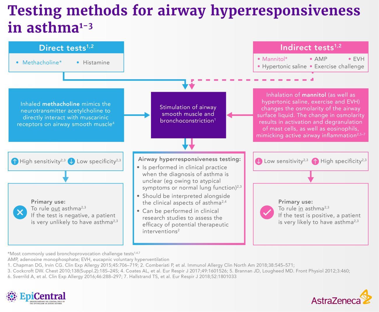
How can airway hyperresponsiveness be measured?

Airway hyperresponsiveness can be measured to confirm or exclude a diagnosis of asthma, classify the severity of asthma or monitor asthma control.3,50–53 Airway hyperresponsiveness can be measured directly or indirectly and involves stimulation of airway smooth muscle with various agents.1,3,54 The two most common methods for testing airway hyperresponsiveness are methacholine, a direct challenge, and mannitol, an indirect challenge.1,3,50,55

Image
Summary of the testing methods used for airway hyperresponsiveness in asthma.

Summary of the testing methods used for airway hyperresponsiveness in asthma.

Click here for a deep dive into the tests for airway hyperresponsiveness, including mechanisms of testing, interpretation of results and contraindications.

The importance of airway hyperresponsiveness in asthma

Airway hyperresponsiveness is a cardinal feature of asthma.1 It is the result of the intricate interplay of airway inflammation, airway remodelling and structural changes, and can be present in the absence of significant airway inflammation.1–3 Understanding the mechanisms of airway hyperresponsiveness can provide greater insights into asthma pathophysiology.1

Find out more about the EpiCreators – Professor Bruce Levy and Professor Chris Brightling

The content for this module was also created with the support of Professor Teal Hallstrand

RELATED RESOURCES

Like this page? We have a host of other resources available in the ‘Scientific and Resource Library’ where you can find out more.

References

1. Chapman DG, Irvin CG. Clin Exp Allergy 2015;45:706–719, 2. Berair R, et al. J Allergy (Cairo) 2013;2013:185971, 3. Comberiati P, et al. Immunol Allergy Clin North Am 2018;38:545–571, 4. Borak J, Lefkowitz RY. Occup Med (Lond) 2016;66:95–105, 5. Allakhverdi Z, et al. J Allergy Clin Immunol 2009;123:958–960, 6. Gunst SJ, Panettieri RA. J Appl Physiol (1985) 2012;113:837–839, 7. Porsbjerg CM, et al. Eur Respir J 2020;56:2000260, 8. Busse WW. Chest 2010;138(Suppl. 2):4S-10S, 9. Crimi E, et al. Am J Respir Crit Care Med 1998;157:4–9, 10. Jeffery PK, et al. Am Rev Respir Dis 1989;140:1745–1753, 11. Boulet LP, et al. Chest 1997;112:45–52, 12. Booms P, et al. J Allergy Clin Immunol 1997;99:330–337, 13. Gelb AF, Zamel N. Curr Opin Pulm Med 2002;8:50–53, 14. Slats AM, et al. J Allergy Clin Immunol 2008;121:1196–1202, 15. Ward C, et al. Thorax 2002;57:309–316, 16. Heijink IH, et al. Allergy 2020;75:1902–1917, 17. Fehrenbach H, et al. Cell Tissue Res 2017;367:551–569, 18. Hough KP, et al. Front Med 2020;7:191, 19. Gil FR, Lauzon A-M. Can J Physiol Pharmacol 2007;85:133–140, 20. Bradding P. Eur Respir J 2007;29:827–830, 21. Hollins F, et al. J Immunol 2008;181:2772–2780, 22. John AE, et al. J Immunol 2009;183:4682–4692, 23. Kaur D, et al. J Immunol 2010;185:6105–6114, 24. Moiseeva EP, et al. PLoS One 2013;8:e61579, 25. Kaur D, et al. Chest 2012;142:76–85, 26. Suto W, et al. Int J Mol Sci 2018;19:3036, 27. Robinson DS. J Allergy Clin Immunol 2004;114:58–65, 28. Brightling CE, et al. N Engl J Med 2002;346:1699–1705, 29. Kaur D, et al. Allergy 2015;70:556–567, 30. Moir LM, et al. J Allergy Clin Immunol 2008;121:1034–1039, 31. Woodman L, et al. J Immunol 2008;181:5001–5007, 32. Tatler AL, et al. J Immunol 2011;187:6094–6107, 33. Saunders R, et al. J Allergy Clin Immunol 2009;123:376–384, 34. Saunders R, et al. Clin Transl Immunology 2020;9:e1205, 35. Singh SR, et al. Allergy 2014;69:1189–1197, 36. Begueret H, et al. Thorax 2007;62:8–15, 37. Siddiqui S, et al. J Allergy Clin Immunol 2008;122:335–341, 38. Bonvini SJ, et al. Eur Respir J 2020;56:1901458, 39. Lai Y, et al. J Allergy Clin Immunol 2014;133:1448–1455, 40. Altman MC, et al. J Clin Invest 2019;129:4979–4991, 41. Al-Shaikhly T, et al. Eur Respir J 2022;60:2101865, 42. Rayees S, Din I. Asthma: pathophysiology, herbal and modern therapeutic interventions. Cham, Switzerland: Springer International Publishing, 2021, 43. in’t Veen JC, et al. Am J Respir Crit Care Med 1999;160:93–99, 44. Leuppi JD, et al. Am J Respir Crit Care Med 2001;163:406–412, 45. Rijcken B, Weiss ST. Am J Respir Crit Care Med 1996;154:S246-249, 46. Sears MR, et al. N Engl J Med 2003;349:1414–1422, 47. Cockcroft DW, et al. Clin Allergy 1977;7:235–243, 48. Boulet LP, et al. J Allergy Clin Immunol 1983;71:399–406, 49. Reddel HK, et al. Eur Respir J 2000;16:226–235, 50. Coates AL, et al. Eur Respir J 2017;49:1601526, 51. Fowler SJ, et al. Am J Respir Crit Care Med 2000;162:1318–1322, 52. Vandenplas O, et al. Eur Respir J 2014;43:1573–1587, 53. Weiler JM, et al. J Allergy Clin Immunol 2016;138:1292–1295, 54. O’Byrne PM, Inman MD. Chest 2003;123:411S-6S, 55. Hallstrand TS, et al. Eur Respir J 2018;52:1801033